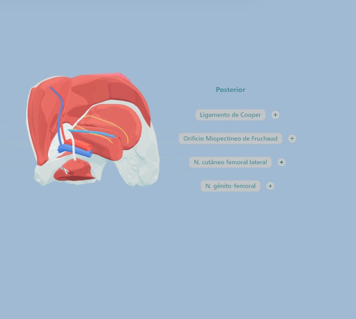
Meet the 3D platform developed by HerniaBrasil for study, teaching, and patient communication.
The anatomy of the inguinal region is not always easy to understand. Transforming two-dimensional images into a three-dimensional view requires time, repetition, and experience. It was with this challenge in mind that we developed a free and open-access 3D anatomical model of the inguinal region, available in Portuguese, English, and Spanish for surgeons worldwide.
The platform allows for a dynamic and intuitive exploration of anatomy. It is possible to rotate the pelvis in any direction, zoom in or out of the view, add or remove structures, and build knowledge progressively. Muscles, ligaments, nerves, vessels, and surgical spaces can be analyzed individually, and upon selecting each element, the user finds objective explanations about its anatomy and clinical relevance.
The idea stemmed from a conviction that guides our daily practice: anatomy is the foundation of safe surgery. Knowing the anatomical planes, recognizing risk areas, and understanding the relationships between structures is fundamental to avoiding complications and making safer decisions in the operating room.
We, Dr. Natália Pascotini Pereira and Dr. Paulo Henrique Fogaça de Barros, are surgeons dedicated to the treatment of abdominal wall hernias and deeply involved in surgical education. This same philosophy guides The Lab – Cadaver Lab: Anatomical Dissection of Hernia Surgery, an immersive course where small groups of surgeons explore anatomy on fresh cadavers, discuss technical details, and learn strategies to make surgery more precise and safe.
The 3D model was born as a digital extension of this commitment to education. Developed in partnership with designer Italo Marques Pessoa, it expands access to anatomical knowledge and allows concepts to be revisited whenever necessary, whether for studying, preparing a lecture, or facilitating patient understanding during a consultation.
Explore the platform, incorporate it into your routine, and share it with other colleagues.
Because when we understand anatomy better, we operate better. And those who benefit most from this are our patients.